Datoteka:Unctional complex and pinocytotic vesicles - embryonic brain - TEM.jpg

Izvorna datoteka (1.600 × 1.278 piksela, veličina datoteke: 861 KB, MIME tip: image/jpeg)
| Ova datoteka je s Wikimedia Commonsa. Opis s njene stranice opisa datoteke prikazan je ispod. Commons je skladište slobodnih medija i datoteka za sve projekte fondacije Wikimedia. Možete i Vi pomoći. |
Sažetak
| Opis |
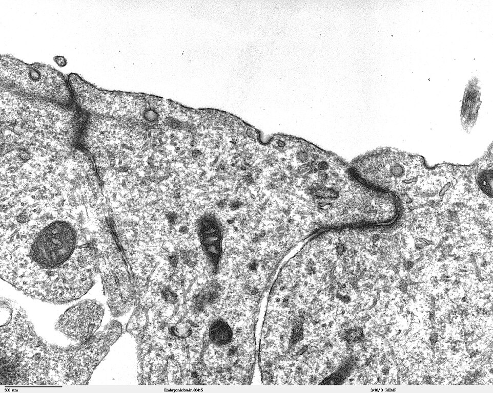
Transmission electron microscope image of a thin section cut through the developing brain tissue (telencephalic hemisphere) of an 11.5 day mouse embryo. This image of the luminal surface of the telencephalon, shows junctional complexes and pinocytotic vesicles. The junctional complex is divided into three types of junctions: 1) the most apical is the tight junction, which controls and/or restricts the movement of molecules across epithelial layers and helps maintain polarity, 2) the zonula adherens, which also includes the numerous actin filaments seen in the apical cytoplasm, and 3) the desmosome, which is a spot junction. The pinocytotic vesicles are formed from coated pits in the plasma membrane and are involved in endocytosis. JEOL 100CX TEM References: Marin-Padilla, M. (1985) "Early Vascularization of the Embryonic Cerebral Cortex: Golgi and Electron Microscope Studies", J. Comparative Neurology, 241:237-249 Marin-Padilla, M. and M. Amievo (1989) "Early Neurogenesis of the Mouse Olfactory Nerve: Golgi and Electron Microscope Studies", J. Comparative Neurology, 288:339-352 |
| Izvor | |
| Autor | Louisa Howard, Miguel Marin-Padilla |
| Dopuštenje (Naknadno korištenje ove datoteke) |
PD |
Licenciranje
| Ovaj rad je objavljen u javno vlasništvo od strane autora Louisa Howard, Miguel Marin-Padilla. Ovo se primjenjuje širom svijeta. U nekim državama ovo zakonski nije moguće; u tom slučaju: Louisa Howard, Miguel Marin-Padilla dopušta svima pravo korištenja ovog rada u bilo koju svrhu, bez ikakvih uslova, osim ako su takvi uslovi zakonski neophodni.
|
Historija datoteke
Kliknite na datum/vrijeme da vidite verziju datoteke iz tog vremena.
| Datum/vrijeme | Smanjeni pregled | Dimenzije | Korisnik | Komentar | |
|---|---|---|---|---|---|
| trenutno | 23:06, 2 novembar 2006 | | 1.600 × 1.278 (861 KB) | Patho | {{Information |Description=Transmission electron microscope image of a thin section cut through the developing brain tissue (telencephalic hemisphere) of an 11.5 day mouse embryo. This higher magnification image of "Embryonic brain 80415", shows an area o |
Upotreba datoteke
Sljedeća stranica koristi ovu datoteku:
Globalna upotreba datoteke
Sljedeći wikiji koriste ovu datoteku:
- Upotreba na ca.wikipedia.org
- Upotreba na de.wikipedia.org
- Upotreba na de.wikibooks.org
- Upotreba na en.wikibooks.org
- Upotreba na et.wikipedia.org
- Upotreba na fr.wikipedia.org