Datoteka:Development of the neural tube.png
Veća rezolucija nije dostupna.
Development_of_the_neural_tube.png (598 × 368 piksela, veličina datoteke: 36 KB, MIME tip: image/png)
| Ova datoteka je s Wikimedia Commonsa. Opis s njene stranice opisa datoteke prikazan je ispod. Commons je skladište slobodnih medija i datoteka za sve projekte fondacije Wikimedia. Možete i Vi pomoći. |
Sažetak
| Opis |
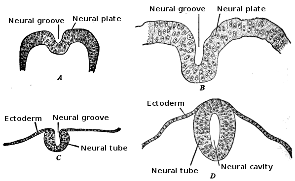
English: Development of the neural tube in human embryos (Prentiss-Arey). A. An early embryo (Keibel) B. at 2 mm. (Graf Spee) C. at 2 mm. (Mall) D. at 2.7 mm (Kollmann).
This is a scan of Figure 6 of the book "The anatomy of the nervous system" by Stephen Walter Ranson, with the labels redrawn. |
||||||||||||||||
| Datum | file created 2009-12-24, original image published 1920 | ||||||||||||||||
| Izvor |
Figure 6 (p. 24) of "The anatomy of the nervous system" by Stephen Walter Ranson, published W.B. Saunders, 1920
|
||||||||||||||||
| Autor | user:Looie496 created file, original artist unknown | ||||||||||||||||
| Ostale verzije |
|
||||||||||||||||

== Licenciranje ==
This media file is in the public domain in the United States. This applies to U.S. works where the copyright has expired, often because its first publication occurred prior to January 1, 1929, and if not then due to lack of notice or renewal. See this page for further explanation.
|
 | |
 |
This image might not be in the public domain outside of the United States; this especially applies in the countries and areas that do not apply the rule of the shorter term for US works, such as Canada, Mainland China (not Hong Kong or Macao), Germany, Mexico, and Switzerland. The creator and year of publication are essential information and must be provided. See Wikipedia:Public domain and Wikipedia:Copyrights for more details.
|
Historija datoteke
Kliknite na datum/vrijeme da vidite verziju datoteke iz tog vremena.
| Datum/vrijeme | Smanjeni pregled | Dimenzije | Korisnik | Komentar | |
|---|---|---|---|---|---|
| trenutno | 22:08, 5 januar 2010 | | 598 × 368 (36 KB) | Looie496 | {{Information |Description={{en|1=Development of the neural tube in human embryos (Prentiss-Arey). A. An early embryo (Keibel) B. at 2 mm. (Graf Spee) C. at 2 mm. (Mall) D. at 2.7 mm (Kollmann). This is a scan of Figure 6 of the book "The anatomy of |
Upotreba datoteke
Sljedeća stranica koristi ovu datoteku:
Globalna upotreba datoteke
Sljedeći wikiji koriste ovu datoteku:
- Upotreba na af.wikipedia.org
- Upotreba na ar.wikipedia.org
- Upotreba na az.wikipedia.org
- Upotreba na bg.wikipedia.org
- Upotreba na en.wikipedia.org
- Upotreba na fr.wikipedia.org
- Upotreba na gl.wikipedia.org
- Upotreba na hr.wikipedia.org
- Upotreba na id.wikipedia.org
- Upotreba na sr.wikipedia.org